Differential Pulmonary Effects of CoO and La2O3 Metal Oxide Nanoparticle Responses during Aerosolized Inhalation in Mice
Supporting Files
Public Domain

-
2016/08/15
-
File Language:
English
Details
-
Journal Article:Particle and Fibre Toxicology
-
Personal Author:Sisler, Jennifer D. ; Li, Ruibin ; McKinney, Walter G. ; Mercer, Robert R. ; Ji, Zhaoxia ; Xia, Tian ; Wang, Xiang ; Shaffer, Justine ; Orandle, Marlene S. ; Mihalchik, Amy L. ; Battelli, Lori A. ; Chen, Bean T. ; Wolfarth, Michael G. ; Andrew, Michael E. ; Schwegler-Berry, Diane ; Porter, Dale W. ; Castranova, Vincent ; Nel, Andre ; Qian, Yong
-
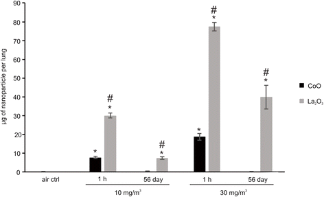
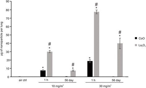
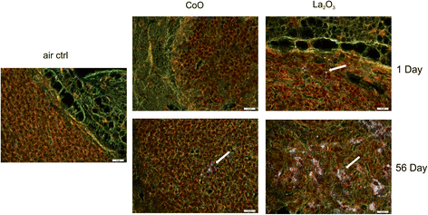
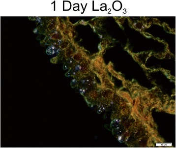
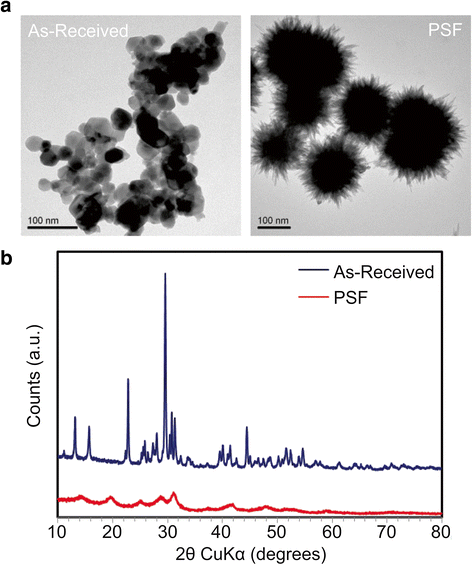
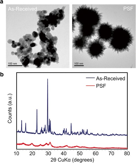
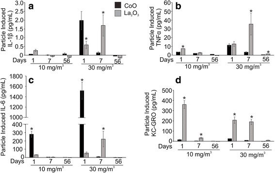
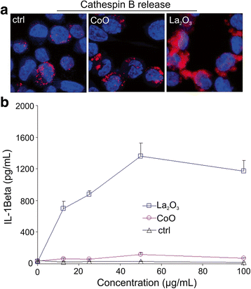
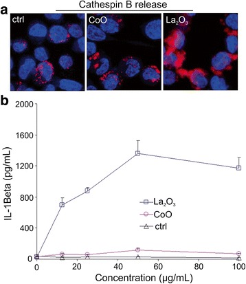
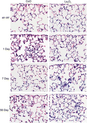
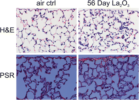
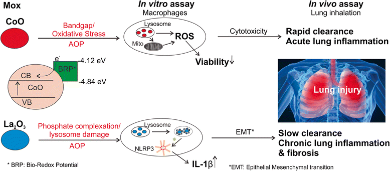
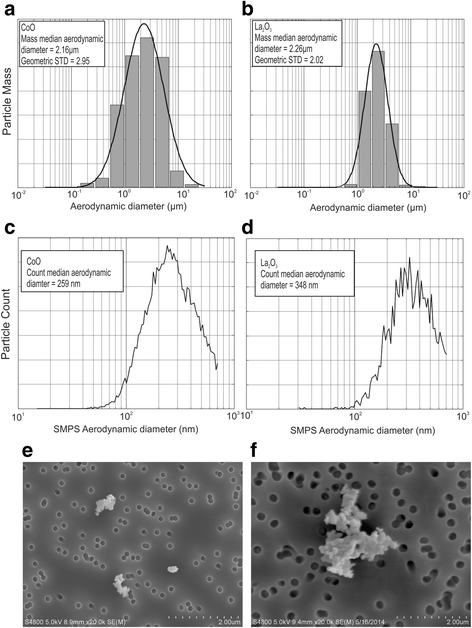
Description:Background: Although classified as metal oxides, cobalt monoxide (CoO) and lanthanum oxide (La2O3) nanoparticles, as representative transition and rare earth oxides, exhibit distinct material properties that may result in different hazardous potential in the lung. The current study was undertaken to compare the pulmonary effects of aerosolized whole body inhalation of these nanoparticles in mice. Results: Mice were exposed to filtered air (control) and 10 or 30 mg/m3 of each particle type for 4 days and then examined at 1 h, 1, 7 and 56 days post-exposure. The whole lung burden 1 h after the 4 day inhalation of CoO nanoparticles was 25 % of that for La2O3 nanoparticles. At 56 days post exposure, < 1 % of CoO nanoparticles remained in the lungs; however, 22-50 % of the La2O3 nanoparticles lung burden 1 h post exposure was retained at 56 days post exposure for low and high exposures. Significant accumulation of La2O3 nanoparticles in the tracheobronchial lymph nodes was noted at 56 days post exposure. When exposed to phagolysosomal simulated fluid, La nanoparticles formed urchin-shaped LaPO4 structures, suggesting that retention of this rare earth oxide nanoparticle may be due to complexation of cellular phosphates within lysosomes. CoO nanoparticles caused greater lactate dehydrogenase release in the bronchoalveolar fluid (BALF) compared to La2O3 nanoparticles at 1 day post exposure, while BAL cell differentials indicate that La2O3 nanoparticles generated more inflammatory cell infiltration at all doses and exposure points. Histopathological analysis showed acute inflammatory changes at 1 day after inhalation of either CoO or La2O3 nanoparticles. Only the 30 mg/m3 La2O3 nanoparticles exposure caused chronic inflammatory changes and minimal fibrosis at day 56 post exposure. This is in agreement with activation of the NRLP3 inflammasome after in vitro exposure of differentiated THP-1 macrophages to La2O3 but not after CoO nanoparticles exposure. Conclusion: Taken together, the inhalation studies confirmed the trend of our previous sub-acute aspiration study, which reported that CoO nanoparticles induced more acute pulmonary toxicity, while La2O3 nanoparticles caused chronic inflammatory changes and minimal fibrosis. [Description provided by NIOSH]
-
Subjects:
-
Keywords:Author Keywords: Nanoparticles; Metal Oxides; Pulmonary Response; In Vivo; Mouse Toxicology; Nanotechnology; Nanoparticles; Rare Earth Metals; Metal Oxides; Lanthanum Oxide; Cobalt Oxides; Lung Irritants; Pulmonary Effects; Respiratory Irritants; Aerosol Particles; Aerosols; Inhalation; Inhalation Studies; Laboratory Animals; Laboratory Testing; Exposure Assessment; Exposure Levels; Lung Burden; Lymph Nodes; Cellular Structures; Chemical Properties; Chemical Structure; Physical Properties; Phosphates; Enzymes; Alveolar Cells; Dose Response; Histopathology; Fibrosis; Lung Fibrosis; Chronic Inflammation; In Vivo Study; Macrophages;
-
Source:Part Fibre Toxicol 2016 Aug; 13:42
-
ISSN:1743-8977
-
Pubmed ID:27527840
-
Pubmed Central ID:PMC4986387
-
Document Type:
-
Genre:
-
Place as Subject:
-
CIO:
-
Division:
-
Topic:
-
Location:
-
Pages in Document:17 pdf pages
-
Volume:13
-
NIOSHTIC Number:nn:20048595
-
Contact Point Address:Yong Qian, Health Effects Laboratory Division, National Institute for Occupational Safety and Health, 1095 Willowdale Road, Morgantown, WV 26505, USA
-
Email:yaq2@cdc.gov
-
CAS Registry Number:
-
Federal Fiscal Year:2016
-
NORA Priority Area:
-
Peer Reviewed:True
-
Collection(s):
-
Main Document Checksum:urn:sha-512:826b0e5e239a068afe897af965814042891e2848b8f8d3f73b0454756df71d12da689c5550288a8532d54824b3ea34063a183682e308e40e12320957ae6f62e5
-
Download URL:
-
File Type:
 [PDF
- 2.97 MB
]
[PDF
- 2.97 MB
]
Supporting Files

File Language:
English
ON THIS PAGE

CDC STACKS serves as an archival repository of CDC-published products including
scientific findings,
journal articles, guidelines, recommendations, or other public health information authored or
co-authored by CDC or funded partners.
As a repository, CDC STACKS retains documents in their original published format to ensure public access to scientific information.
As a repository, CDC STACKS retains documents in their original published format to ensure public access to scientific information.
You May Also Like