Chronic fetal hypoxia affects axonal maturation in guinea pigs during development: a longitudinal Diffusion Tensor Imaging and T2 mapping study
Supporting Files

-
Dec 15 2014
-
Details
-
Alternative Title:J Magn Reson Imaging
-
Personal Author:
-
Description:Purpose
Chronic hypoxemia is the prime cause of fetal brain injury and long-term sequelae such as neurodevelopmental compromise, seizures and cerebral palsy. This study aims to investigate the impact of chronic hypoxemia on neonatal brains, and follow developmental alterations and adaptations non-invasively in a guinea pig model.
Materials and Methods
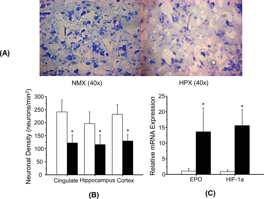
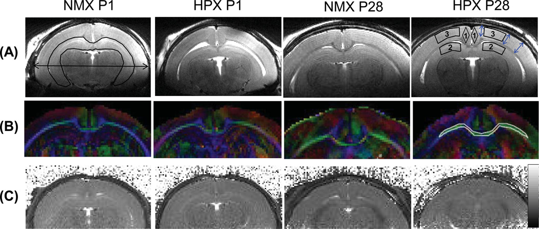
Thirty guinea pigs underwent either normoxic and hypoxemic conditions during the critical stage of brain development (0.7 gestation) and studied prenatally (n=16) or perinatally (n=14). Fourteen newborns (7 hypoxia and 7 normoxia group) were scanned longitudinally to characterize physiological and morphological alterations, and axonal myelination and injury using in vivo DTI, T2 mapping, and T2-weighted MRI. Sixteen fetuses (8 hypoxia and 8 normoxia) were studied ex vivo to assess hypoxia-induced neuronal injury/loss using Nissl staining and quantitative reverse transcriptase Polymerase Chain Reaction methods.
Results
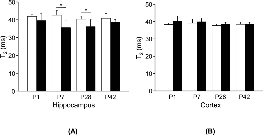
Developmental brains in the hypoxia group showed lower fractional anisotropy in the corpus callosum (−12%, p=0.02) and lower T2 values in the hippocampus (−16%, p=0.003) compared with the normoxia group with no differences in the cortex (p>0.07), indicating vulnerability of the hippocampus and cerebral white matter during early development. Fetal guinea pig brains with chronic hypoxia demonstrated an over-tenfold increase in expression levels of hypoxia index genes such as erythropoietin and HIF-1α, and an over 40% reduction in neuronal density, confirming prenatal brain damage.
Conclusion
In vivo MRI measurement, such as DTI and T2 mapping, provides quantitative parameters to characterize neuro-developmental abnormalities and to monitor the impact of prenatal insult on the postnatal brain maturation of guinea pigs.
-
Subjects:
-
Source:J Magn Reson Imaging. 42(3):658-665.
-
Pubmed ID:25504885
-
Pubmed Central ID:PMC4468050
-
Document Type:
-
Funding:DP00187-5/DP/NCCDPHP CDC HHS/United States ; P30 AG035982/AG/NIA NIH HHS/United States ; P30 HD002528/HD/NICHD NIH HHS/United States ; R01 HL049041/HL/NHLBI NIH HHS/United States ; R01 HL049041-13/HL/NHLBI NIH HHS/United States ; R03 HD062734/HD/NICHD NIH HHS/United States ; S10 RR029577/RR/NCRR NIH HHS/United States ; S10 RR29577/RR/NCRR NIH HHS/United States ; UL1 RR033179/RR/NCRR NIH HHS/United States ; UL1RR033179/RR/NCRR NIH HHS/United States
-
Volume:42
-
Issue:3
-
Collection(s):
-
Main Document Checksum:urn:sha256:22768e8131ff30eebe6fc76a76522595aff2fd8f91e2d979ca2891fd1f920f6f
-
Download URL:
-
File Type:
 [PDF
- 1.71 MB
]
[PDF
- 1.71 MB
]
Supporting Files

ON THIS PAGE

CDC STACKS serves as an archival repository of CDC-published products including
scientific findings,
journal articles, guidelines, recommendations, or other public health information authored or
co-authored by CDC or funded partners.
As a repository, CDC STACKS retains documents in their original published format to ensure public access to scientific information.
As a repository, CDC STACKS retains documents in their original published format to ensure public access to scientific information.
You May Also Like
COLLECTION
CDC Public Access