Expansion Microscopy
Supporting Files

-
Jan 15 2015
Details
-
Alternative Title:Science
-
Personal Author:
-
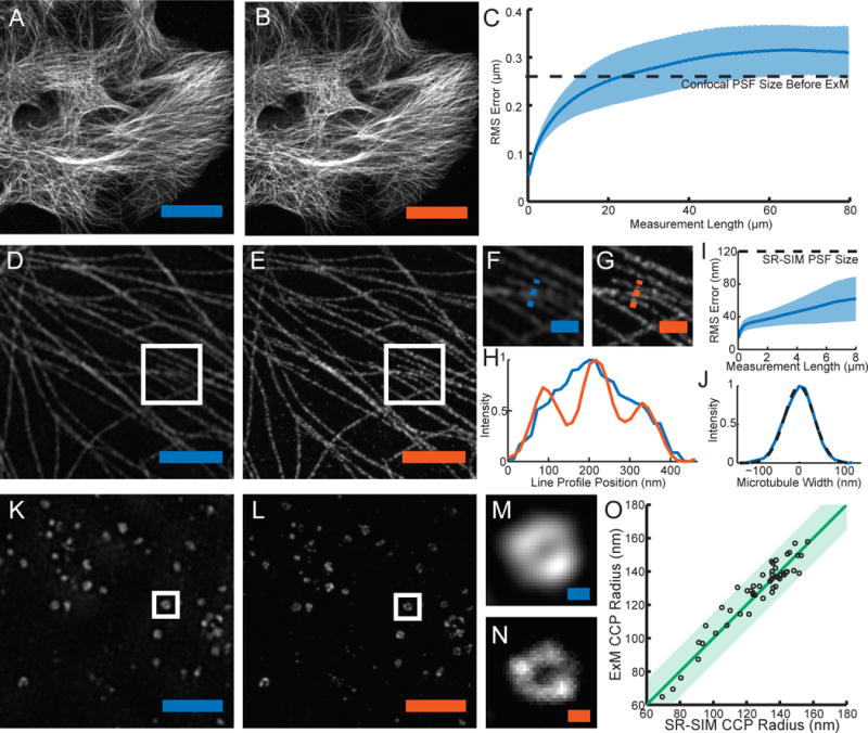
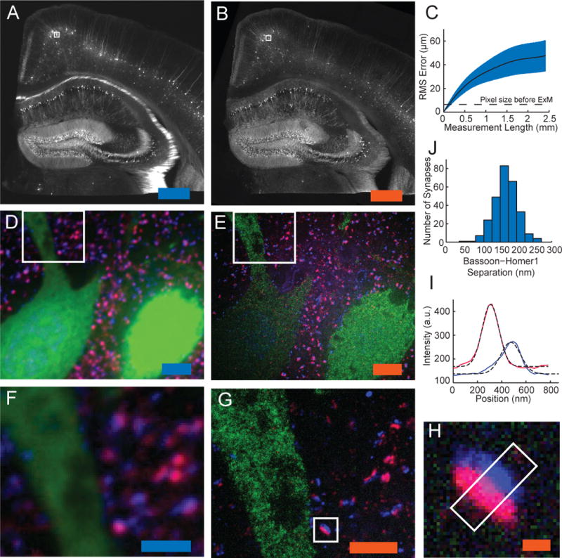
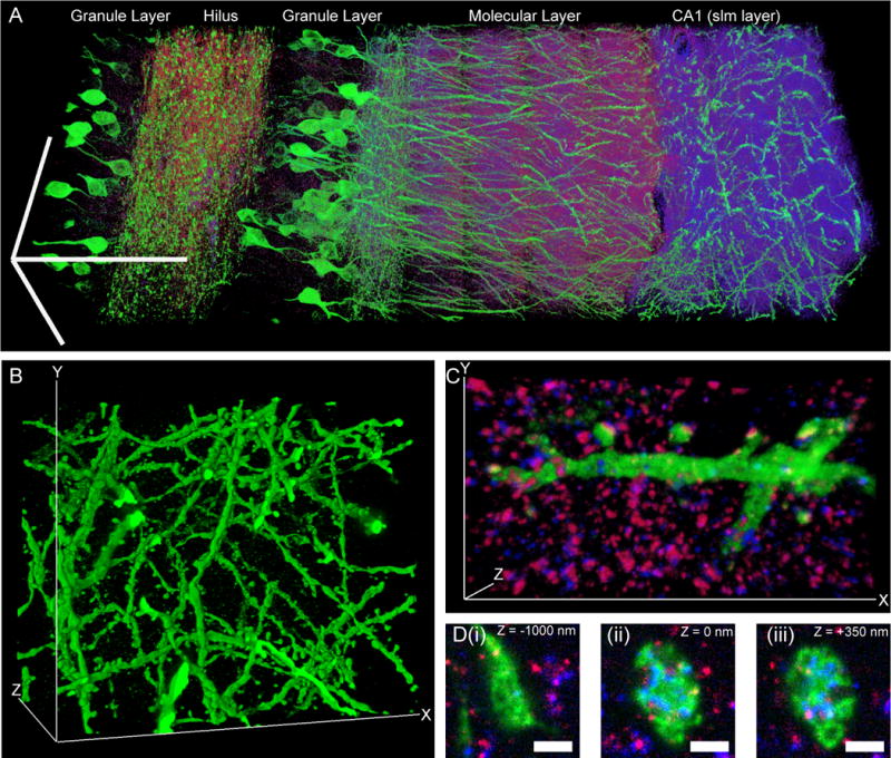
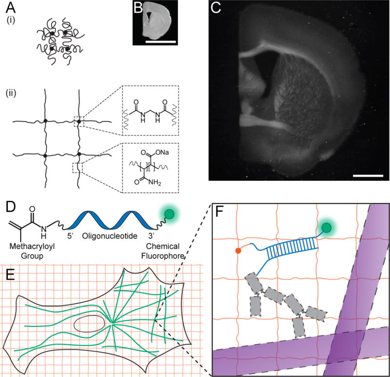
Description:In optical microscopy, fine structural details are resolved by using refraction to magnify images of a specimen. We discovered that by synthesizing a swellable polymer network within a specimen, it can be physically expanded, resulting in physical magnification. By covalently anchoring specific labels located within the specimen directly to the polymer network, labels spaced closer than the optical diffraction limit can be isotropically separated and optically resolved, a process we call expansion microscopy (ExM). Thus, this process can be used to perform scalable superresolution microscopy with diffraction-limited microscopes. We demonstrate ExM with apparent ~70-nanometer lateral resolution in both cultured cells and brain tissue, performing three-color superresolution imaging of ~10(7) cubic micrometers of the mouse hippocampus with a conventional confocal microscope.
-
Subjects:
-
Source:Science. 347(6221):543-548.
-
Pubmed ID:25592419
-
Pubmed Central ID:PMC4312537
-
Document Type:
-
Funding:
-
Volume:347
-
Issue:6221
-
Collection(s):
-
Main Document Checksum:urn:sha256:6e059be7ee19365a37efd59f9ce92a35338dbdb931af4438c5bfd543ec3226fd
-
Download URL:
-
File Type:
 [PDF
- 1013.63 KB
]
[PDF
- 1013.63 KB
]
Supporting Files

ON THIS PAGE

CDC STACKS serves as an archival repository of CDC-published products including
scientific findings,
journal articles, guidelines, recommendations, or other public health information authored or
co-authored by CDC or funded partners.
As a repository, CDC STACKS retains documents in their original published format to ensure public access to scientific information.
As a repository, CDC STACKS retains documents in their original published format to ensure public access to scientific information.
You May Also Like
COLLECTION
CDC Public Access