Cardiac R2* values are independent on the image analysis approach employed
Supporting Files

-
October 01 2013
-
File Language:
English
Details
-
Alternative Title:Magn Reson Med
-
Personal Author:
-
Description:Purpose
To determine whether systematic differences were present between myocardial R2* values obtained with two different decay models: truncation and exponential-plus-constant (Exp-C).
Methods
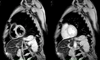
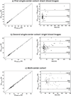
Single-center cohorts were used to compare black and bright blood sequences separately and a multi-center cohort of mixed bright and black blood studies was used to assess the generalizability. Truncated exponential estimates were calculated with CMRTools that uses a single region of interest (ROI) method. Exp-C estimates were calculated using a pixelwise approach.
Results
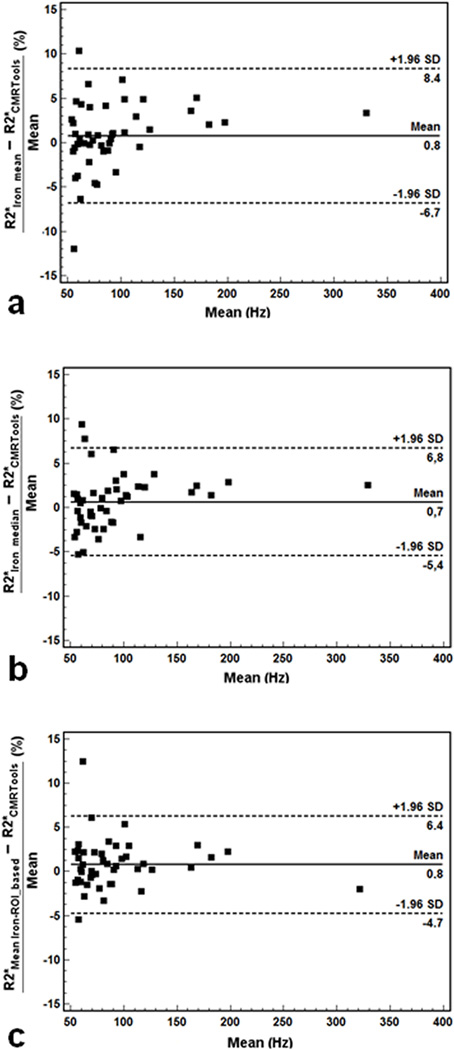
No differences could be distinguished based upon whether a white or black blood sequence was examined. The two fitting algorithms gave similar R2* values, with R-squared values exceeding 0.997 and CoV of 3–4%. Results using the pixelwise method yielded a small systematic bias (~3%) that became apparent in patients with severe iron deposition. This disparity disappeared when Exp-C fitting was used on a single ROI suggesting that the use of pixelwise mapping was responsible for the bias. In the multicenter cohort the strong agreement between the two fitting approaches was reconfirmed.
Conclusion
Cardiac R2* values are independent of the signal model used for its calculation over clinically relevant ranges. Clinicians can compare results among centers using these disparate approaches with confidence.
-
Subjects:
-
Source:Magn Reson Med. 72(2):485-491
-
Pubmed ID:24123261
-
Pubmed Central ID:PMC4293021
-
Document Type:
-
Funding:RC1 HL099412/HL/NHLBI NIH HHS/United States ; RR00043-43/RR/NCRR NIH HHS/United States ; 1 U01 DD000309-1/DD/NCBDD CDC HHS/United States ; R01 HL075592/HL/NHLBI NIH HHS/United States ; M01 RR000043/RR/NCRR NIH HHS/United States ; U01 DD000309/DD/NCBDD CDC HHS/United States ; 1 R01 HL075592-01A1/HL/NHLBI NIH HHS/United States
-
Volume:72
-
Issue:2
-
Collection(s):
-
Main Document Checksum:urn:sha256:b708fc1a53367bda2c4b8b9db9da6a0caa01fb37ff134965c3f6e91ed27c55b8
-
Download URL:
-
File Type:
 [PDF
- 789.23 KB
]
[PDF
- 789.23 KB
]
Supporting Files

File Language:
English
ON THIS PAGE

CDC STACKS serves as an archival repository of CDC-published products including
scientific findings,
journal articles, guidelines, recommendations, or other public health information authored or
co-authored by CDC or funded partners.
As a repository, CDC STACKS retains documents in their original published format to ensure public access to scientific information.
As a repository, CDC STACKS retains documents in their original published format to ensure public access to scientific information.
You May Also Like
COLLECTION
CDC Public Access