Routine argyrophil techniques detect Rickettsia rickettsii in tissues of patients with fatal Rocky Mountain spotted fever
Supporting Files

-
2016
-
File Language:
English
Details
-
Journal Article:J Histotechnol
-
Personal Author:
-
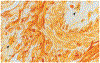
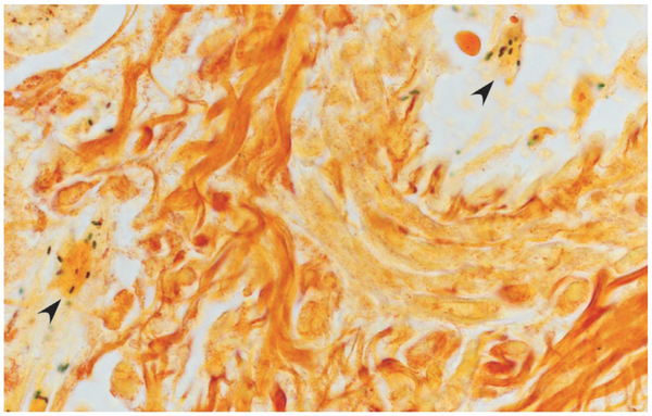
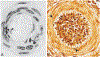
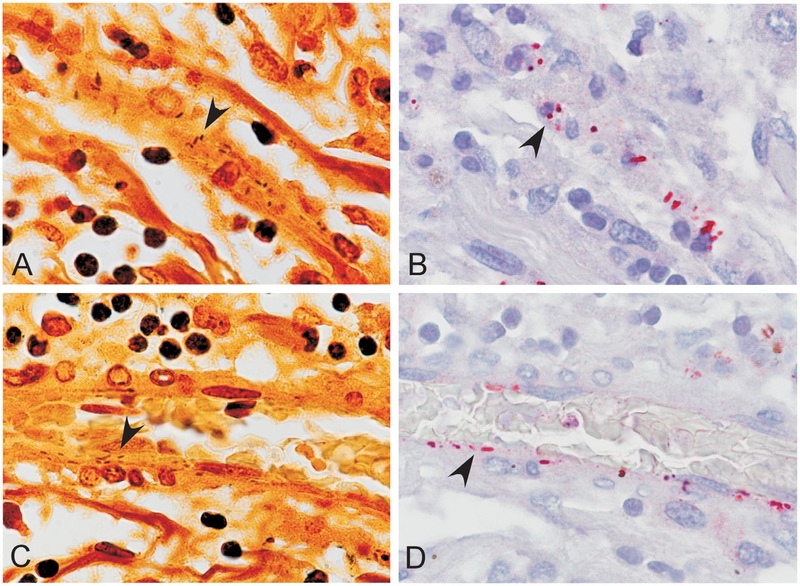
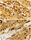
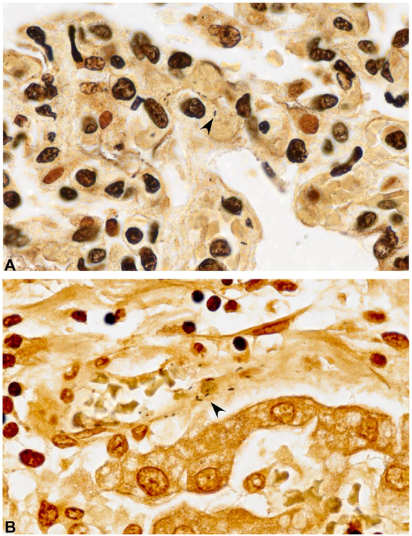
Description:, a bacterial tickborne pathogen that causes Rocky Mountain spotted fever (RMSF), stains poorly or not at all with conventional tissue Gram techniques, and contemporary visualization of the pathogen in formalin-fixed, paraffin-embedded tissues has relied almost entirely on immunohistochemical staining methods that are generally limited to specialized research laboratories or national reference centers. To our knowledge, previously described argyrophil-based histochemical techniques have not successfully detected rickettsiae in formalin-fixed, paraffin-embedded tissues. To investigate the ability of standard silver impregnation techniques to demonstrate the occurrence and distribution of | in tissues of patients with RMSF confirmed by molecular and immunohistochemical methods, three widely recognized and commercially available silver impregnation methods (Warthin-Starry, Steiner, and Dieterle's) were applied to various tissues obtained at autopsy from 10 patients with fatal RMSF. | bacteria were demonstrated in one or more tissues of all patients, using each of the argyrophil-based methods, and appeared as small, dark brown-to-black lanceolate rods, often in pairs and occasionally surrounded by a faint halo. Rickettsiae were identified most consistently in small arteries and arterioles of liver, kidney, and leptomeninges, and were localized predominantly to the cytoplasm of endothelial cells and less often within the internal elastic lamella and smooth muscle of the media. This validation of argyrophilic techniques to detect | demonstrates the utility of inexpensive core histochemical methods in the diagnosis of infectious agents in pathology specimens and may have utility in certain resource-limited settings where RMSF is endemic.
-
Keywords:
-
Source:J Histotechnol. 39(4):116-122
-
DOI:
-
Pubmed ID:32636574
-
Pubmed Central ID:PMC7340092
-
Document Type:
-
Funding:
-
Genre:
-
Volume:39
-
Issue:4
-
Collection(s):
-
Main Document Checksum:urn:sha-512:13f913a1d3b834c9ad9983549c5f5c8624fbd071fe57b4a7f623c0563e6e372f49603dd540ed97ce5f1f6f83825237b54b376be1cc1c0e1cbe274e1d3afd02dc
-
Download URL:
-
File Type:
 [PDF
- 1.96 MB
]
[PDF
- 1.96 MB
]
Supporting Files

File Language:
English
ON THIS PAGE

CDC STACKS serves as an archival repository of CDC-published products including
scientific findings,
journal articles, guidelines, recommendations, or other public health information authored or
co-authored by CDC or funded partners.
As a repository, CDC STACKS retains documents in their original published format to ensure public access to scientific information.
As a repository, CDC STACKS retains documents in their original published format to ensure public access to scientific information.
You May Also Like
COLLECTION
CDC Public Access