Clinical, Histopathologic, and Immunohistochemical Characterization of Experimental Marburg Virus Infection in A Natural Reservoir Host, the Egyptian Rousette Bat (Rousettus aegyptiacus)
Supporting Files

-
March 02 2019
-
File Language:
English
Details
-
Journal Article:Viruses
-
Personal Author:
-
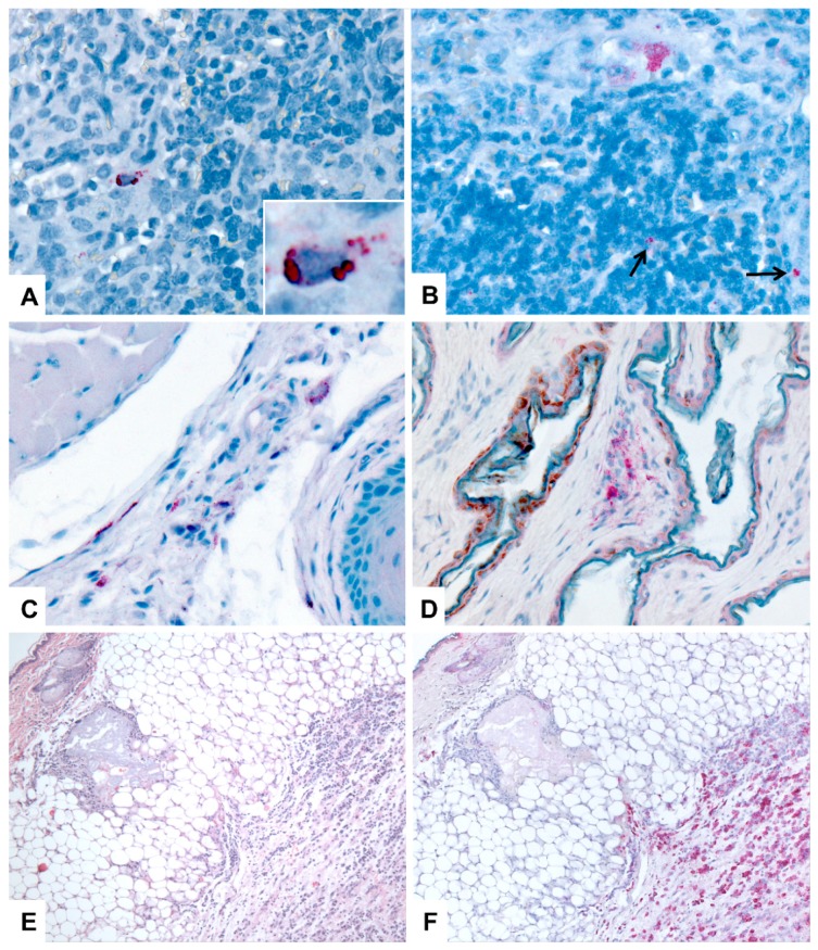
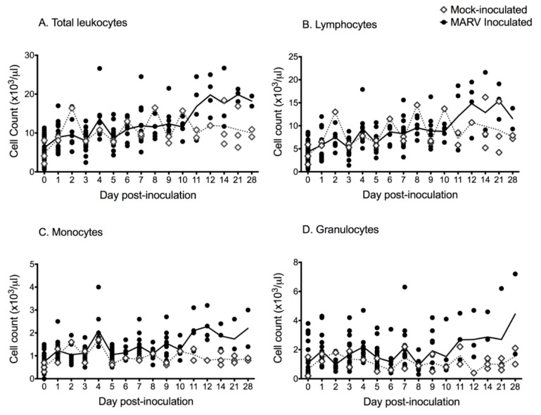
Description:Egyptian rousette bats (|) are natural reservoir hosts of Marburg virus (MARV), and Ravn virus (RAVV; collectively called marburgviruses) and have been linked to human cases of Marburg virus disease (MVD). We investigated the clinical and pathologic effects of experimental MARV infection in Egyptian rousettes through a serial euthanasia study and found clear evidence of mild but transient disease. Three groups of nine, captive-born, juvenile male bats were inoculated subcutaneously with 10,000 TCID| of Marburg virus strain Uganda 371Bat2007, a minimally passaged virus originally isolated from a wild Egyptian rousette. Control bats (| = 3) were mock-inoculated. Three animals per day were euthanized at 3, 5⁻10, 12 and 28 days post-inoculation (DPI); controls were euthanized at 28 DPI. Blood chemistry analyses showed a mild, statistically significant elevation in alanine aminotransferase (ALT) at 3, 6 and 7 DPI. Lymphocyte and monocyte counts were mildly elevated in inoculated bats after 9 DPI. Liver histology revealed small foci of inflammatory infiltrate in infected bats, similar to lesions previously described in wild, naturally-infected bats. Liver lesion severity scores peaked at 7 DPI, and were correlated with both ALT and hepatic viral RNA levels. Immunohistochemical staining detected infrequent viral antigen in liver (3⁻8 DPI, | = 8), spleen (3⁻7 DPI, | = 8), skin (inoculation site; 3⁻12 DPI, | = 20), lymph nodes (3⁻10 DPI, | = 6), and oral submucosa (8⁻9 DPI, | = 2). Viral antigen was present in histiocytes, hepatocytes and mesenchymal cells, and in the liver, antigen staining co-localized with inflammatory foci. These results show the first clear evidence of very mild disease caused by a filovirus in a reservoir bat host and provide support for our experimental model of this virus-reservoir host system.
-
Subjects:
-
Source:Viruses. 2019; 11(3)
-
DOI:
-
Pubmed ID:30832364
-
Pubmed Central ID:PMC6466277
-
Document Type:
-
Funding:
-
Genre:
-
Volume:11
-
Issue:3
-
Collection(s):
-
Main Document Checksum:urn:sha-512:64bdf7a6475a76901ee9604e7565d0998ad8ab4e6b340802279b668393d73ba20b3ac48943afa763b20c9c14743b36386e29a1802bba641ee971faae5519bdfc
-
Download URL:
-
File Type:
 [PDF
- 2.64 MB
]
[PDF
- 2.64 MB
]
Supporting Files

File Language:
English
ON THIS PAGE

CDC STACKS serves as an archival repository of CDC-published products including
scientific findings,
journal articles, guidelines, recommendations, or other public health information authored or
co-authored by CDC or funded partners.
As a repository, CDC STACKS retains documents in their original published format to ensure public access to scientific information.
As a repository, CDC STACKS retains documents in their original published format to ensure public access to scientific information.
You May Also Like
COLLECTION
CDC Public Access