Photoacoustic tomography imaging and estimation of oxygen saturation of hemoglobin in ocular tissue of rabbits
Supporting Files

-
9 2015
-
File Language:
English
Details
-
Alternative Title:Exp Eye Res
-
Personal Author:
-
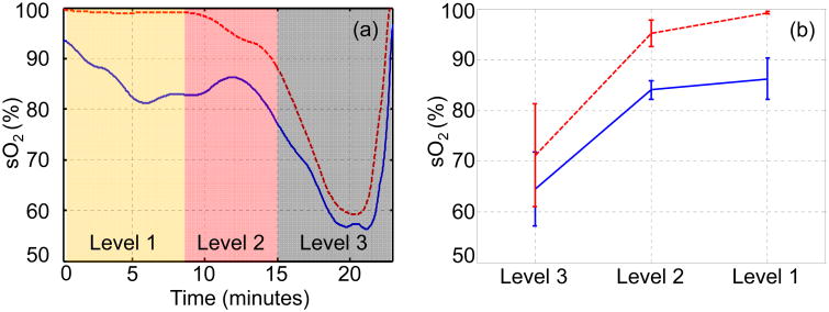
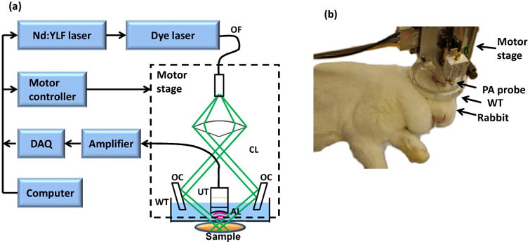
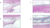
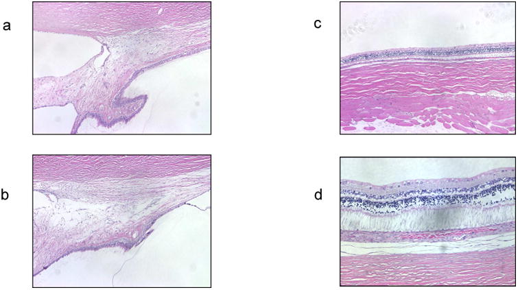
Description:This study evaluated in vivo imaging capabilities and safety of qualitative monitoring of oxygen saturation of hemoglobin (sO2) of rabbit ciliary body tissues obtained with acoustic resolution (AR) photoacoustic tomography (PAT). AR PAT was used to collect trans-scleral images from ciliary body vasculature of seven New Zealand White rabbits. The PAT sO2 measurements were obtained under the following conditions: when systemic sO2 as measured by pulse oximetry was between 100% and 99% (level 1); systemic sO2 as measured by pulse oximetry was between 98% and 90% (level 2); and systemic sO2 as measured by pulse oximetry was less than 90% (level 3). Following imaging, histological analysis of ocular tissue was conducted to evaluate for possible structural damage caused by the AR PAT imaging. AR PAT was able to resolve anatomical structures of the anterior segment of the eye, viewed through the cornea or anterior sclera. Histological studies revealed no ocular damage. On average, sO2 values (%) obtained with AR PAT were lower than sO2 values obtained with pulse oximetry (all p < 0.001): 86.28 ± 4.16 versus 99.25 ± 0.28, 84.09 ± 1.81 vs. 95.3 ± 2.6, and 64.49 ± 7.27 vs. 71.15 ± 10.21 for levels 1, 2 and 3 respectively. AR PAT imaging modality is capable of qualitative monitoring for deep tissue sO2 in rabbits. Further studies are needed to validate and modify the AR PAT modality specifically for use in human eyes. Having a safe, non-invasive method of in vivo imaging of sO2 in the anterior segment is important to studies evaluating the role of oxidative damage, hypoxia and ischemia in pathogenesis of ocular diseases.
-
Keywords:
-
Source:Exp Eye Res. 138:153-158
-
Pubmed ID:26048477
-
Pubmed Central ID:PMC5821107
-
Document Type:
-
Funding:
-
Volume:138
-
Collection(s):
-
Main Document Checksum:urn:sha256:5e551a73ad1f694086661c4600d6888dc5550e0275932f1ebae2014daca185b8
-
Download URL:
-
File Type:
 [PDF
- 651.09 KB
]
[PDF
- 651.09 KB
]
Supporting Files

File Language:
English
ON THIS PAGE

CDC STACKS serves as an archival repository of CDC-published products including
scientific findings,
journal articles, guidelines, recommendations, or other public health information authored or
co-authored by CDC or funded partners.
As a repository, CDC STACKS retains documents in their original published format to ensure public access to scientific information.
As a repository, CDC STACKS retains documents in their original published format to ensure public access to scientific information.
You May Also Like
COLLECTION
CDC Public Access