Vascular-Directed Responses of Microglia Produced by Methamphetamine Exposure: Indirect Evidence that Microglia Are Involved in Vascular Repair?
Supporting Files
Public Domain

-
2016/03/12
-
File Language:
English
Details
-
Journal Article:Journal of Neuroinflammation
-
Personal Author:
-
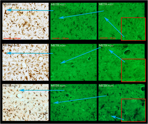
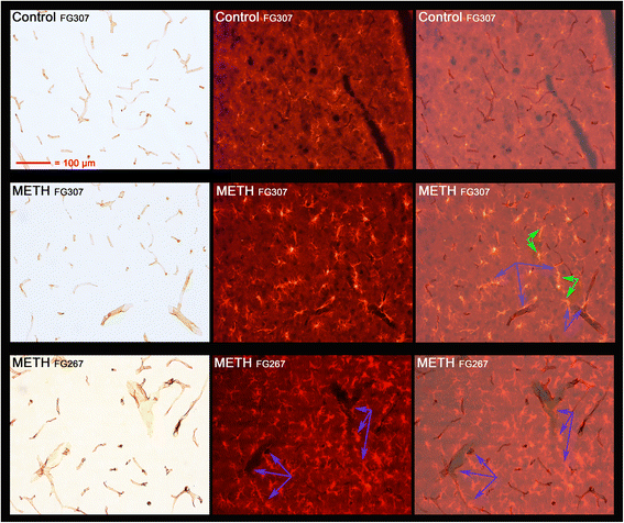
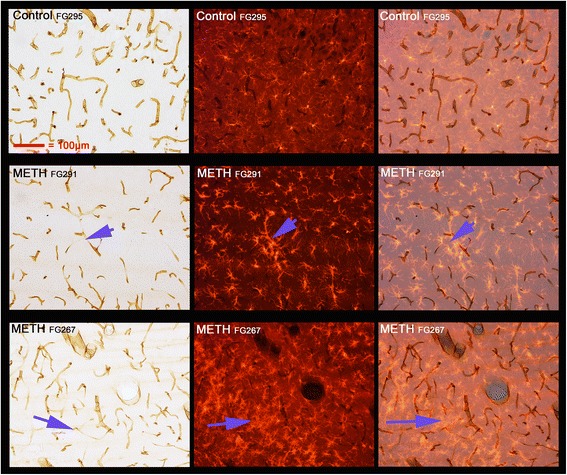
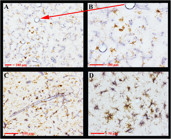
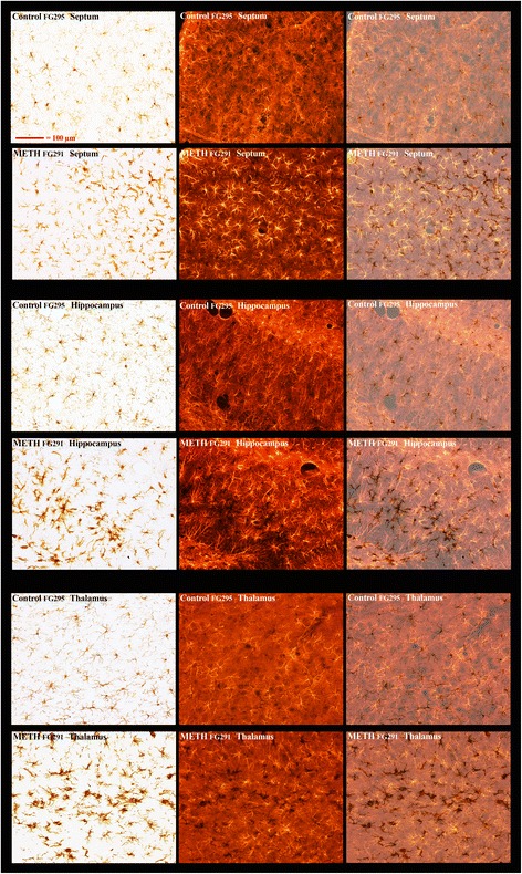
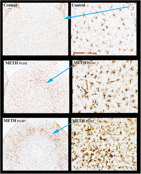
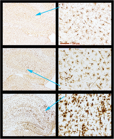
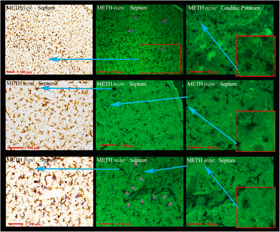
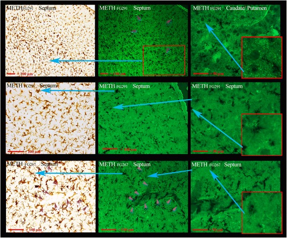
Description:Background: Brain microglial activations and damage responses are most commonly associated with neurodegeneration or systemic innate immune system activation. Here, we used histological methods to focus on microglial responses that are directed towards brain vasculature, previously undescribed, after a neurotoxic exposure to methamphetamine. Methods: Male rats were given doses of methamphetamine that produce pronounced hyperthermia, hypertension, and toxicity. Identification of microglia and microglia-like cells (pericytes and possibly perivascular cells) was done using immunoreactivity to allograft inflammatory factor 1 (Aif1 a.k.a Iba1) and alpha M integrin (Itgam a.k.a. Cd11b) while vasculature endothelium was identified using rat endothelial cell antigen 1 (RECA-1). Regions of neuronal, axonal, and nerve terminal degeneration were determined using Fluoro-Jade C. Results: Dual labeling of vasculature (RECA-1) and microglia (Iba1) showed a strong association of hypertrophied cells surrounding and juxtaposed to vasculature in the septum, medial dorsal hippocampus, piriform cortex, and thalamus. The Iba1 labeling was more pronounced in the cell body while Cd11b more so in the processes of activated microglia. These regions have been previously identified to have vascular leakage after neurotoxic methamphetamine exposure. Dual labeling with Fluoro-Jade C and Iba1 indicated that there was minimal or no evidence of neuronal damage in the septum and hippocampus where many hypertrophied Iba1-labeled cells were found to be associated with vasculature. Although microglial activation around the prominent neurodegeneration was found in the thalamus, there were also many examples of activated microglia associated with vasculature. Conclusions: The data implicate microglia, and possibly related cell types, in playing a major role in responding to methamphetamine-induced vascular damage, and possibly repair, in the absence of neurodegeneration. Identifying brain regions with hypertrophied/activated microglial-like cells associated with vasculature has the potential for identifying regions of more subtle examples of vascular damage and BBB compromise. [Description provided by NIOSH]
-
Subjects:
-
Keywords:Author Keywords: Microglia; Vascular Damage; Methamphetamine; Amphetamine; Neurotoxicity; Hyperthermia; Hypertension Neurological System; Brain Function; Neurotoxic Effects; Neurotoxicity; Exposure Levels; Risk Factors; Methamphetamine; Animals; Laboratory Animals; Hyperthermia; Hypertension; Toxic Effects; Endothelial Cells;
-
Source:J Neuroinflammation 2016 Mar; 13(1):64
-
ISSN:1742-2094
-
Pubmed ID:26970737
-
Pubmed Central ID:PMC4789274
-
Document Type:
-
Genre:
-
Place as Subject:
-
CIO:
-
Division:
-
Topic:
-
Location:
-
Pages in Document:15 pdf pages
-
Volume:13
-
Issue:1
-
NIOSHTIC Number:nn:20047754
-
Contact Point Address:John F. Bowyer, Division of Neurotoxicology, National Center for Toxicology/FDA, Jefferson, AR 72079
-
Email:John.Bowyer@fda.hhs.gov
-
Federal Fiscal Year:2016
-
Peer Reviewed:True
-
Collection(s):
-
Main Document Checksum:urn:sha-512:1b22b9fbb64a25d833ef8d12cbe0db53d3c9ced27ef5f3e9196f852a6964039e1cd2f484f8828b796164a83bb1d2cd7b4ca0f4be96ccb4c7bd2e99a35b3f91cb
-
Download URL:
-
File Type:
 [PDF
- 7.38 MB
]
[PDF
- 7.38 MB
]
Supporting Files

File Language:
English
ON THIS PAGE

CDC STACKS serves as an archival repository of CDC-published products including
scientific findings,
journal articles, guidelines, recommendations, or other public health information authored or
co-authored by CDC or funded partners.
As a repository, CDC STACKS retains documents in their original published format to ensure public access to scientific information.
As a repository, CDC STACKS retains documents in their original published format to ensure public access to scientific information.
You May Also Like