In Vivo Time-gated Fluorescence Imaging with Biodegradable Luminescent Porous Silicon Nanoparticles
Supporting Files

-
2013
-
File Language:
English
Details
-
Alternative Title:Nat Commun
-
Personal Author:
-
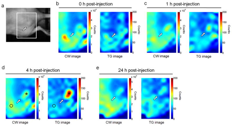
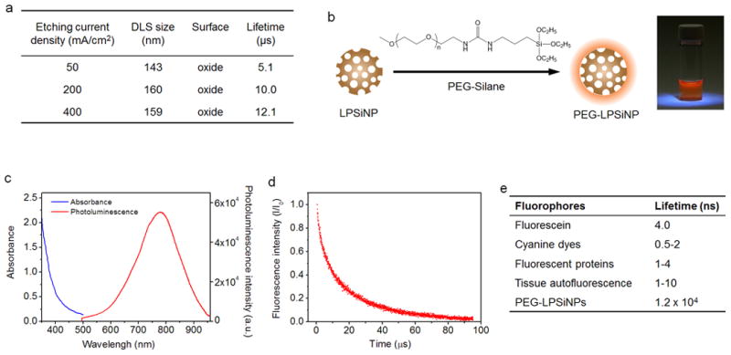
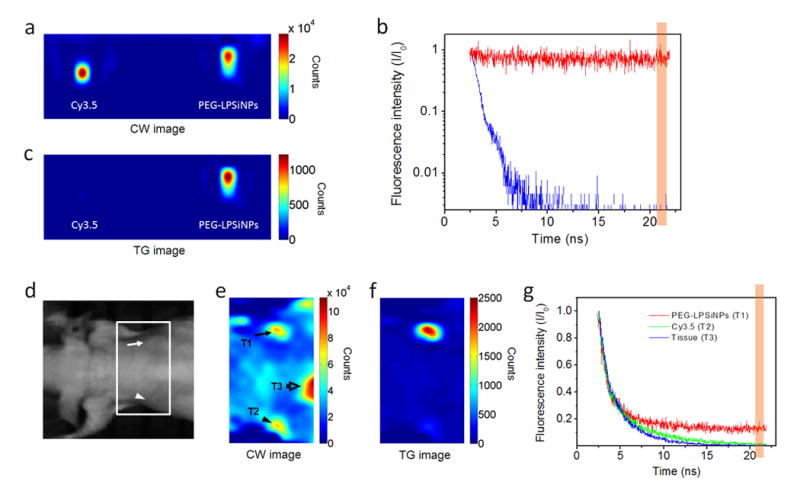
Description:Fluorescence imaging is one of the most versatile and widely used visualization methods in biomedical research. However, tissue autofluorescence is a major obstacle confounding interpretation of in vivo fluorescence images. The unusually long emission lifetime (5-13 μs) of photoluminescent porous silicon nanoparticles can allow the time-gated imaging of tissues in vivo, completely eliminating shorter-lived (<10 ns) emission signals from organic chromophores or tissue autofluorescence. Here using a conventional animal imaging system not optimized for such long-lived excited states, we demonstrate improvement of signal to background contrast ratio by >50-fold in vitro and by >20-fold in vivo when imaging porous silicon nanoparticles. Time-gated imaging of porous silicon nanoparticles accumulated in a human ovarian cancer xenograft following intravenous injection is demonstrated in a live mouse. The potential for multiplexing of images in the time domain by using separate porous silicon nanoparticles engineered with different excited state lifetimes is discussed.
-
Subjects:
-
Source:Nat Commun. 4:2326
-
Pubmed ID:23933660
-
Pubmed Central ID:PMC4154512
-
Document Type:
-
Funding:CA095298/CA/NCI NIH HHS/United States ; U54 CA119335/CA/NCI NIH HHS/United States ; U54-CA 119335/CA/NCI NIH HHS/United States ; R01 CA152185/CA/NCI NIH HHS/United States ; 5R01EB015498/EB/NIBIB NIH HHS/United States ; CA152185/CA/NCI NIH HHS/United States ; R01 CA095298/CA/NCI NIH HHS/United States ; DP2 OD006499/OD/NIH HHS/United States ; 1DP20D006499-01/DP/NCCDPHP CDC HHS/United States ; HHMI56005681/Howard Hughes Medical Institute/United States ; P50CA128346/CA/NCI NIH HHS/United States ; Howard Hughes Medical Institute/United States ; R01 EB015498/EB/NIBIB NIH HHS/United States ; 5-R01-CA124427/CA/NCI NIH HHS/United States ; P50 CA128346/CA/NCI NIH HHS/United States ; R01 CA124427/CA/NCI NIH HHS/United States
-
Volume:4
-
Collection(s):
-
Main Document Checksum:urn:sha256:a3bdc1fc9bc21ad9c570d11994c868dbf5dbdada0fc41378d6b60fe09aed089c
-
Download URL:
-
File Type:
 [PDF
- 498.50 KB
]
[PDF
- 498.50 KB
]
Supporting Files

File Language:
English
ON THIS PAGE

CDC STACKS serves as an archival repository of CDC-published products including
scientific findings,
journal articles, guidelines, recommendations, or other public health information authored or
co-authored by CDC or funded partners.
As a repository, CDC STACKS retains documents in their original published format to ensure public access to scientific information.
As a repository, CDC STACKS retains documents in their original published format to ensure public access to scientific information.
You May Also Like
COLLECTION
CDC Public Access