Use of Contrast-Enhanced Sonography to Investigate Intraneural Vascularity in a Cohort of Macaca fascicularis With Suspected Median Mononeuropathy
Supporting Files

-
Jan 2014
-
Details
-
Alternative Title:J Ultrasound Med
-
Personal Author:
-
Description:Objectives
The purpose of this study was to provide clinical evidence of the use of contrast-enhanced sonography in detecting and quantifying changes in intraneural vascularity due to median mononeuropathy.
Methods
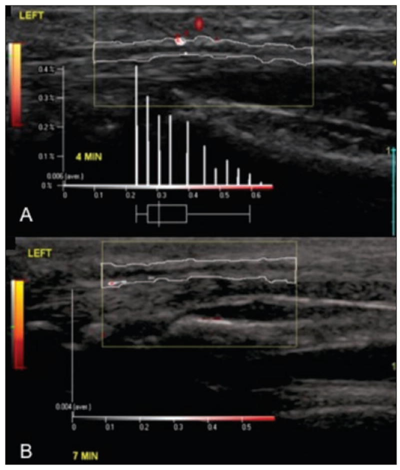
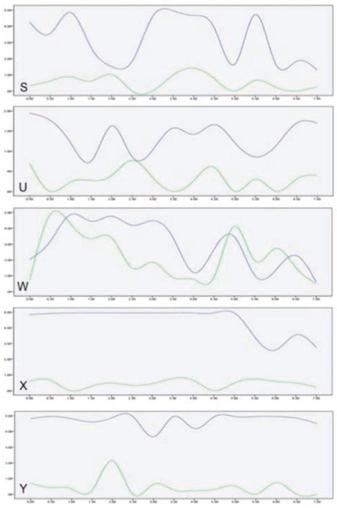
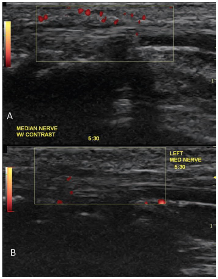
Five Macaca fascicularis monkeys were exposed to 20 weeks of repetitive work to increase their risk of developing median mononeuropathy. Contrast-enhanced sonograms were obtained in 30-second increments for 7 minutes while a contrast agent was being delivered. Data were collected immediately at the conclusion of the 20-week work exposure and then again during a recovery phase approximately 3 months after the completion of work. Quantitative analysis and trend graphs were used to analyze median nerve perfusion intensity. This study also compared the use of both manual counting of pixels and semiautomatic measurement using specialized software.
Results
Based on the average data, maximum intensity values were identified as the best indicators of nerve hyperemia. Paired t tests demonstrated significantly higher maximum intensities in the working stage for 4 of the 5 subjects (P < .01).
Conclusions
This study provides preliminary evidence that (1) in a controlled exposure model, a change in intraneural vascularity of the median nerve between working and recovery can be observed; (2) this vascular change can be measured using an objective technique that quantifies the intensity of vascularity; and (3) contrast-enhanced sonography may improve the ability to reliably capture and measure low-flow microvascularity.
-
Subjects:
-
Source:J Ultrasound Med. 2014; 33(1):103-109.
-
Pubmed ID:24371104
-
Pubmed Central ID:PMC4040227
-
Document Type:
-
Funding:UL1 TR001070/TR/NCATS NIH HHS/United States ; 5R21OH009907-02/OH/NIOSH CDC HHS/United States ; UL1RR025755/RR/NCRR NIH HHS/United States ; R21 OH009907/OH/NIOSH CDC HHS/United States ; K12 HD055929/HD/NICHD NIH HHS/United States ; UL1 RR025755/RR/NCRR NIH HHS/United States ; K12HD055929/HD/NICHD NIH HHS/United States
-
Volume:33
-
Issue:1
-
Collection(s):
-
Main Document Checksum:urn:sha256:882343064934e0aa7b5e43f5933fb14bbde455693d29b01bc6a023294526bbfe
-
Download URL:
-
File Type:
 [PDF
- 466.01 KB
]
[PDF
- 466.01 KB
]
Supporting Files

ON THIS PAGE

CDC STACKS serves as an archival repository of CDC-published products including
scientific findings,
journal articles, guidelines, recommendations, or other public health information authored or
co-authored by CDC or funded partners.
As a repository, CDC STACKS retains documents in their original published format to ensure public access to scientific information.
As a repository, CDC STACKS retains documents in their original published format to ensure public access to scientific information.
You May Also Like
COLLECTION
CDC Public Access