Expression of the stem cell marker Nanog in human endometrial adenocarcinoma
Supporting Files

-
5 2011
-
File Language:
English
Details
-
Journal Article:Int J Gynecol Pathol
-
Personal Author:
-
Description:OBJECTIVES
The embryonic stem cell (ESC) self-renewal gene Nanog has been shown to be expressed in several tumor types and to regulate tumor development. The aim of this study was to perform a detailed analysis of Nanog expression in human endometrial adenocarcinoma (EAC).
METHODS
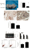
Immunohistochemical analysis and reverse transcriptase-polymerase chain reaction (RT-PCR) were used to characterize Nanog, Sox2, and Oct4 expression in tissue arrays containing EAC, benign endometrium samples, and tumorosphere cells. Tumorosphere formation of EAC-derived cells in stem cell culture medium was also analyzed. Nanog expression was then analyzed in secondary tumors initiated by injection of tumorospheres or tumorosphere-derived differentiated cells into fifteen female nude mice. Apoptosis and cell proliferation were detected in fluorescence activated cell sorter (FACS) and 3-( 4, 5-dimethylthiazolyl-2)-2, 5-diphenyltetrazolium bromide (MTT) experiments, respectively.
RESULTS
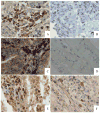
The Nanog protein was expressed in a majority of EAC samples (45/55, 81.8%), but not in benign endometrium samples (0/26, 0.0%). Oct4 and Sox2 were also commonly expressed in EAC samples (42/55, 76.4%; 39/55, 70.9%, respectively). Subsets of cancer cells from all EAC samples (15/15, 100%) exhibited the capacity to form Nanog-positive tumorospheres. The tumorospheres also expressed Nanog, Oct4, and Sox2 mRNA and showed a higher proliferation potential than differentiated cells. All 15 mice that were injected with tumorosphere cells formed tumors, while only 3/15 mice injected with differentiated cells derived from tumorospheres developed tumors. All secondary xenograft tumors still expressed Nanog protein as well as Nanog, Oct4, and Sox2 mRNA, and had higher proliferation and lower apostosis than did differentiated cells.
CONCLUSION
Overexpression of Nanog in EACs suggests that Nanog may represent a potential therapeutic target for EAC. Additionally, Nanog may be useful as a biomarker in an immunohistochemical panel to differentiate between EAC and benign endometrial tissues. Expression of Nanog in tumorospheres may be indicative of the presence of a population of endometrial cancer stem cells (ECSCs), and its expression in xenograft tumors suggests that Nanog may also be associated with tumor metastasis.
-
Subjects:
-
Source:Int J Gynecol Pathol. 30(3):262-270
-
DOI:
-
Pubmed ID:21464727
-
Pubmed Central ID:PMC3077452
-
Document Type:
-
Funding:
-
Genre:
-
Volume:30
-
Issue:3
-
Collection(s):
-
Main Document Checksum:urn:sha-512:a007987a89d3e0e2024fdcb815f04efa3bbc6a6a5d1897cadbe4d957de450b3c592054ec8a282af13fee59f973600146b72da738ee0e26b8858e70c0f1197914
-
Download URL:
-
File Type:
 [PDF
- 495.84 KB
]
[PDF
- 495.84 KB
]
Supporting Files

File Language:
English
ON THIS PAGE

CDC STACKS serves as an archival repository of CDC-published products including
scientific findings,
journal articles, guidelines, recommendations, or other public health information authored or
co-authored by CDC or funded partners.
As a repository, CDC STACKS retains documents in their original published format to ensure public access to scientific information.
As a repository, CDC STACKS retains documents in their original published format to ensure public access to scientific information.
You May Also Like
COLLECTION
CDC Public Access