i
Structures and operating principles of the replisome
-
January 24 2019
-
-
Source: Science. 363(6429)
 [PDF-2.73 MB]
Details:
-
Alternative Title:Science
-
Personal Author:
-
Description:INTRODUCTION
DNA replication has been studied since the 1950s. It is well established that double helical DNA needs to be separated for replication by a helicase. Each strand is then copied by a DNA polymerase, continuously on the leading and discontinuously (via Okazaki fragments) on the lagging strand, where each DNA synthesis initiates from an RNA primer provided by primase. After six decades, how DNA polymerases, helicase, primase, and their accessory factors form a replisome and perform concerted leading and lagging strand synthesis at a replication fork had never been visualized in atomic detail.
RATIONALE
Bacteriophage T7 presents the simplest known DNA replication system, consisting of only three proteins. Helicase and primase reside in one polypeptide chain that forms a hexamer in the presence of DNA and ATP or dTTP. T7 DNA polymerase, aided by E. coli thioredoxin as its processivity factor, carries out both leading and lagging strand DNA synthesis. Based on published biochemical data, we designed a minimal DNA fork to trap these essential proteins in replication competent states.
RESULTS
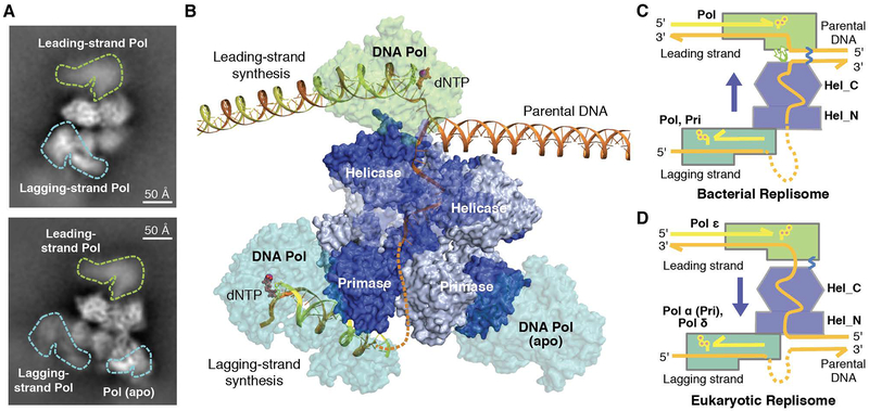
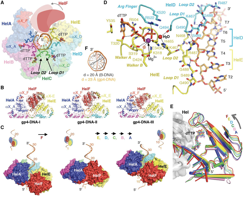
We determined cryogenic-electron microscopy (cryo-EM) structures of the T7 replisome and showed how its essential enzymatic functions are coordinated in three dimensions. The hexameric helicase adopts a spiral “lock washer” form that encircles the coil-like lagging DNA strand, with two nucleotides (nt) bound to each protein subunit and adjacent helicase subunits linked by domain swapping. ATP hydrolysis propels each helicase domain to translocate sequentially and coaxially along DNA in a hand-over-hand fashion, advancing 2 nt per step in the 5′ to 3′ direction (Fig. A). Instead of all enzymes moving in the same direction parallel to the downstream parental DNA, a β-hairpin from the leading-strand polymerase separates the two parental DNA strands into a T-shaped fork that enables the closely coupled helicase to unspool the downstream DNA tangentially (Fig. B). By protein-protein and DNA-mediated interactions, the leading-strand DNA polymerase and helicase cooperate to determine the rate of replication. For every ATP hydrolyzed and 2 nt advanced on DNA by the helicase, the DNA polymerase incorporates two deoxyribonucelotides. T7 primase, separated from the leading-strand polymerase by the helicase domain, synthesizes the RNA primers needed to initiate lagging-strand DNA synthesis. Transfer of a short RNA primer from the primase to DNA polymerase is facilitated by a zinc-binding-domain at the N-terminus of the T7 primase-helicase protein. Two lagging strand polymerases can be attached to the hexameric primases with one actively synthesizing DNA and the other waiting for a primer (Fig. B). Such a relay system may allow the discontinuous lagging-strand synthesis to keep pace with the leading-strand synthesis.
CONCLUSION
We note the similarity between hexameric DNA helicases and AAA+ protein chaperones and unfoldases, which form spiral-shaped hexamers around protein substrates, bind two amino-acid residues with each subunit and move proteins by a hand-over-hand subunit translocation mechanism. The operating principles of the bacteriophage replisome observed here rationalize many well-known features of bacterial and eukaryotic replication. Despite moving in opposite directions (3′ to 5′ on the leading strand in eukaryotes vs. 5′ to 3′ on the lagging strand in bacteria) and association with divergent polymerases, primase and accessory factors, in each replisome a helicase is the central organizer with leading- and lagging-strand DNA synthesis occurring on opposite sides of the helicase. The tight association of leading-strand DNA synthesis with movement of the replication fork sheds light on the effects of DNA lesions on replication and also the direct transfer of histones from parental DNA to the newly synthesized daughter strands in eukaryotes.
-
Subjects:
-
Source:
-
Pubmed ID:30679383
-
Pubmed Central ID:PMC6681829
-
Document Type:
-
Funding:ZIA DK075037-10/Intramural NIH HHS/Intramural NIH HHS/United States ; ZIA DK036146/NIDDK NIH HHS/National Institute of Diabetes and Digestive and Kidney Diseases/United States ; ZIA DK036146-12/Intramural NIH HHS/Intramural NIH HHS/United States ; S10 OD018111/ODCDC CDC HHS/Office of the Director/United States ; U24 GM116792/NIGMS NIH HHS/National Institute of General Medical Sciences/United States ; ... More +
-
Collection(s):
-
Main Document Checksum:
-
Download URL:
-
File Type:


