Serial Head and Brain Imaging of 17 Fetuses With Confirmed Zika Virus Infection in Colombia, South America
Supporting Files

-
7 2017
-
File Language:
English
Details
-
Alternative Title:Obstet Gynecol
-
Personal Author:Parra-Saavedra, Miguel ; Reefhuis, Jennita ; Piraquive, Juan Pablo ; Gilboa, Suzanne M. ; Badell, Martina L. ; Moore, Cynthia A. ; Mercado, Marcela ; Valencia, Diana ; Jamieson, Denise J. ; Beltran, Mauricio ; Sanz-Cortes, Magda ; Rivera-Casas, Ana Maria ; Yepez, Mayel ; Parra, Guido ; Martinez, Martha Ospina ; Honein, Margaret A.
-
Description:Objective
To evaluate fetal ultrasound and magnetic resonance imaging findings among a series of pregnant women with confirmed Zika virus infection to evaluate the signs of congenital Zika syndrome with respect to timing of infection.
Methods
Retrospective case series of pregnant women referred to two perinatal clinics in Barranquilla and Ibagué, Colombia with findings consistent with congenital Zika syndrome and Zika virus infection confirmed in maternal, fetal, or infant samples. Serial ultrasound measurements, fetal magnetic resonance imaging results, laboratory results, and perinatal outcomes were evaluated.
Results
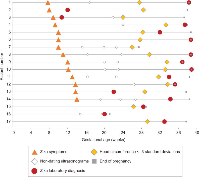
We describe 17 cases of confirmed prenatal maternal Zika virus infection with adverse fetal outcomes. Among the 14 symptomatic women, the median gestational age for maternal Zika virus symptoms was 10 weeks (range 7–14). The median time between Zika virus symptom onset and microcephaly (head circumference less than 3 standard deviations below the mean) was 18 weeks (range 15–24). The earliest fetal head circumference measurement consistent with microcephaly diagnosis was at 24 weeks of gestation. The earliest sign of congenital Zika syndrome was talipes equinovarus, which in two patients was noted first at 19 weeks. Common findings on fetal magnetic resonance imaging were microcephaly, ventriculomegaly, polymicrogyria, and calcifications.
Conclusion
Our analysis suggests a period of at least 15 weeks between maternal Zika virus infection in pregnancy and development of microcephaly, and highlights the importance of serial and detailed neuroimaging.
-
Subjects:
-
Source:Obstet Gynecol. 130(1):207-212
-
Pubmed ID:28594771
-
Pubmed Central ID:PMC5511628
-
Document Type:
-
Funding:
-
Volume:130
-
Issue:1
-
Collection(s):
-
Main Document Checksum:urn:sha256:58f7c26ade9ad71dfaa76d4368ea7287b420d26d8e84de7525f48c369dbacecb
-
Download URL:
-
File Type:
 [PDF
- 341.92 KB
]
[PDF
- 341.92 KB
]
Supporting Files

File Language:
English
ON THIS PAGE

CDC STACKS serves as an archival repository of CDC-published products including
scientific findings,
journal articles, guidelines, recommendations, or other public health information authored or
co-authored by CDC or funded partners.
As a repository, CDC STACKS retains documents in their original published format to ensure public access to scientific information.
As a repository, CDC STACKS retains documents in their original published format to ensure public access to scientific information.
You May Also Like
COLLECTION
CDC Public Access