In Vitro Expansion of Corneal Endothelial Cells on Biomimetic Substrates
Supporting Files

-
Jan 22 2015
-
Details
-
Alternative Title:Sci Rep
-
Personal Author:
-
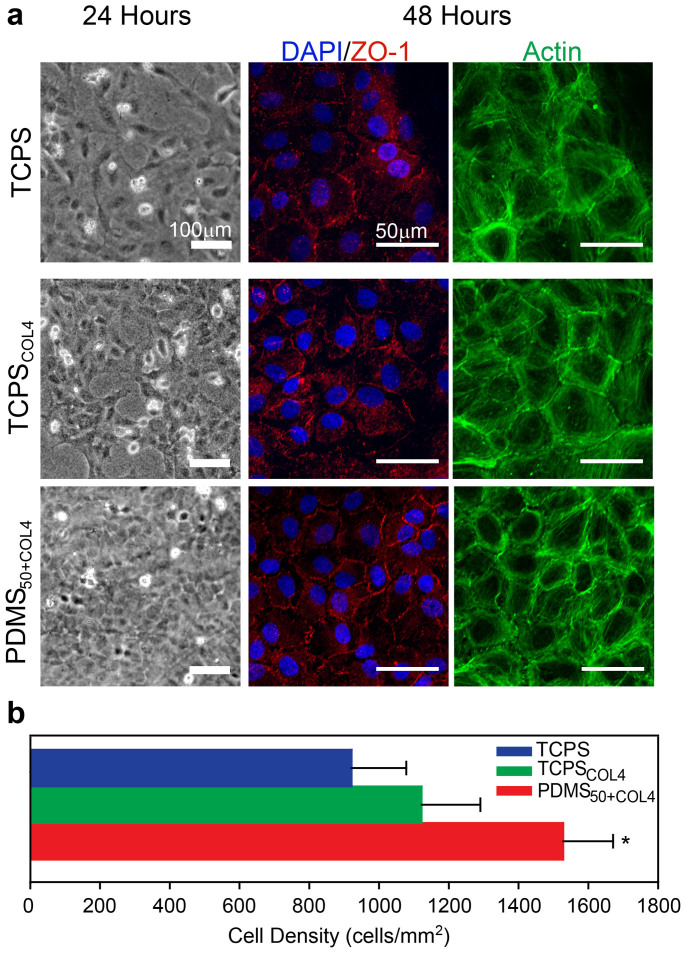
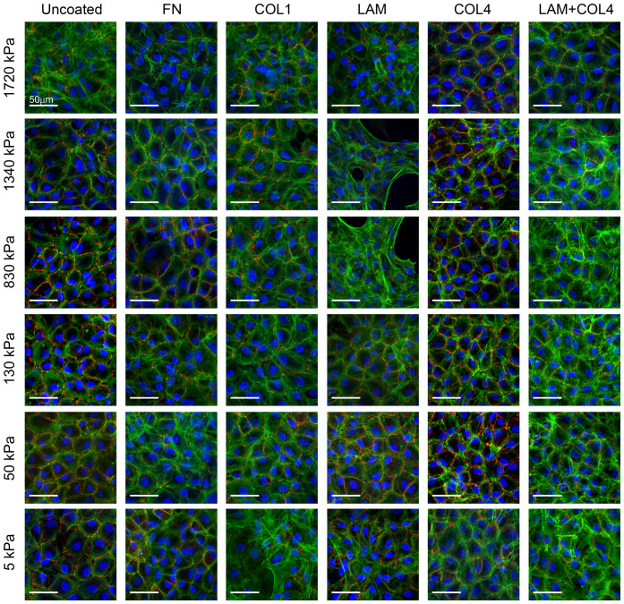
Description:Corneal endothelial (CE) cells do not divide in vivo, leading to edema, corneal clouding and vision loss when the density drops below a critical level. The endothelium can be replaced by transplanting allogeneic tissue; however, access to donated tissue is limited worldwide resulting in critical need for new sources of corneal grafts. In vitro expansion of CE cells is a potential solution, but is challenging due to limited proliferation and loss of phenotype in vitro via endothelial to mesenchymal transformation (EMT) and senescence. We hypothesized that a bioengineered substrate recapitulating chemo-mechanical properties of Descemet's membrane would improve the in vitro expansion of CE cells while maintaining phenotype. Results show that bovine CE cells cultured on a polydimethylsiloxane surface with elastic modulus of 50 kPa and collagen IV coating achieved >3000-fold expansion. Cells grew in higher-density monolayers with polygonal morphology and ZO-1 localization at cell-cell junctions in contrast to control cells on polystyrene that lost these phenotypic markers coupled with increased α-smooth muscle actin expression and fibronectin fibril assembly. In total, these results demonstrate that a biomimetic substrate presenting native basement membrane ECM proteins and mechanical environment may be a key element in bioengineering functional CE layers for potential therapeutic applications.
-
Subjects:
-
Source:Sci Rep. 2015; 5.
-
Pubmed ID:25609008
-
Pubmed Central ID:PMC4302312
-
Document Type:
-
Funding:
-
Volume:5
-
Collection(s):
-
Main Document Checksum:urn:sha256:37521f03bf97772d43b81107d314341f8940b41a7f3475cee3e103aef847c2c5
-
Download URL:
-
File Type:
 [PDF
- 1.65 MB
]
[PDF
- 1.65 MB
]
Supporting Files

ON THIS PAGE

CDC STACKS serves as an archival repository of CDC-published products including
scientific findings,
journal articles, guidelines, recommendations, or other public health information authored or
co-authored by CDC or funded partners.
As a repository, CDC STACKS retains documents in their original published format to ensure public access to scientific information.
As a repository, CDC STACKS retains documents in their original published format to ensure public access to scientific information.
You May Also Like
COLLECTION
CDC Public Access