HISTOLOGIC LESIONS IN PLACENTAS OF NORTHERN FUR SEALS (CALLORHINUS URSINUS) FROM A POPULATION WITH HIGH PLACENTAL PREVALENCE OF COXIELLA BURNETII
Supporting Files

-
4 01 2022
-
File Language:
English
Details
-
Alternative Title:J Wildl Dis
-
Personal Author:
-
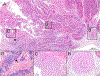
Description:Coxiella burnetii is an intracellular bacterial pathogen that can be associated with significant reproductive disease or acute mortality in livestock and wildlife. A novel marine mammal-associated strain of C. burnetii has been identified in pinnipeds of the northwestern Pacific Ocean. Little is known about C. burnetii infection in regard to reproductive success or population status. Our objective was to characterize the severity and extent of histologic lesions in 117 opportunistically collected placentas from presumed-normal northern fur seals (Callorhinus ursinus) in July 2011 on St. Paul Island, Alaska, US, where a high placental prevalence of C. burnetii had been reported. Sections were examined by histology and immunohistochemistry and impression smears with modified acid-fast stain. The nature and frequency of histologic changes were compared with target COM1 PCR-confirmed C. burnetii positive and negative placentas. Overall, histologic changes were similar to placental lesions described in aborting ruminants; however, changes were variable within and between placentas. Vasculitis and occasional intracellular bacteria were seen only in C. burnetii PCR-positive placentas. Dystrophic mineralization, edema, and inflammation were seen in PCR-positive and negative placentas, although they were statistically more common in PCR-positive placentas. Results suggest that C. burnetti and associated pathologic changes are multifocal and variable in placentas from these presumably live-born pups. Therefore, multiple sections of tissue from different placental areas should be examined microscopically, and screened by PCR, to ensure accurate diagnosis as the genomes per gram of placenta may not necessarily represent the severity of placental disease. These limitations should inform field biologists, diagnosticians, and pathologists how best to screen and sample for pathogens and histopathology in marine mammal placental samples.
-
Subjects:
-
Keywords:
-
Source:J Wildl Dis. 58(2):333-340
-
Pubmed ID:35245373
-
Pubmed Central ID:PMC11290099
-
Document Type:
-
Funding:
-
Volume:58
-
Issue:2
-
Collection(s):
-
Main Document Checksum:urn:sha-512:2ad879878702810491ea4b1f70008c8c817811b2e078484f4227a48eb9268da7714792f7eb0f5bf031f5ff29dec3f8f7bd70b3261d880306c764a5b6f7d82878
-
Download URL:
-
File Type:
 [PDF
- 936.17 KB
]
[PDF
- 936.17 KB
]
Supporting Files

File Language:
English
ON THIS PAGE

CDC STACKS serves as an archival repository of CDC-published products including
scientific findings,
journal articles, guidelines, recommendations, or other public health information authored or
co-authored by CDC or funded partners.
As a repository, CDC STACKS retains documents in their original published format to ensure public access to scientific information.
As a repository, CDC STACKS retains documents in their original published format to ensure public access to scientific information.
You May Also Like
COLLECTION
CDC Public Access