i
Microglial activation and responses to vasculature that result from an acute LPS exposure
-
January 31 2020
Source: Neurotoxicology. 77:181-192 -
Alternative Title:Neurotoxicology
-
Personal Author:
-
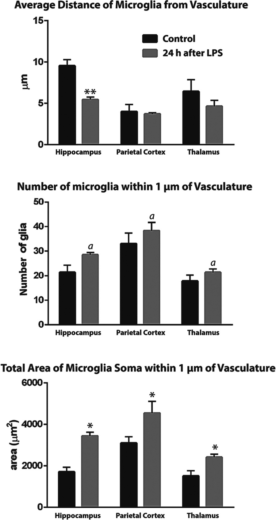
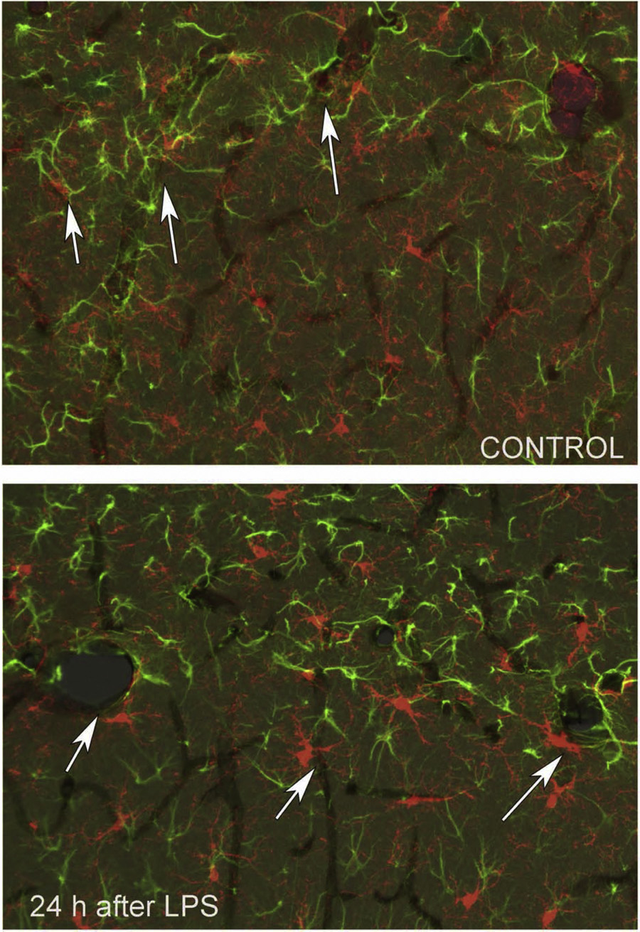
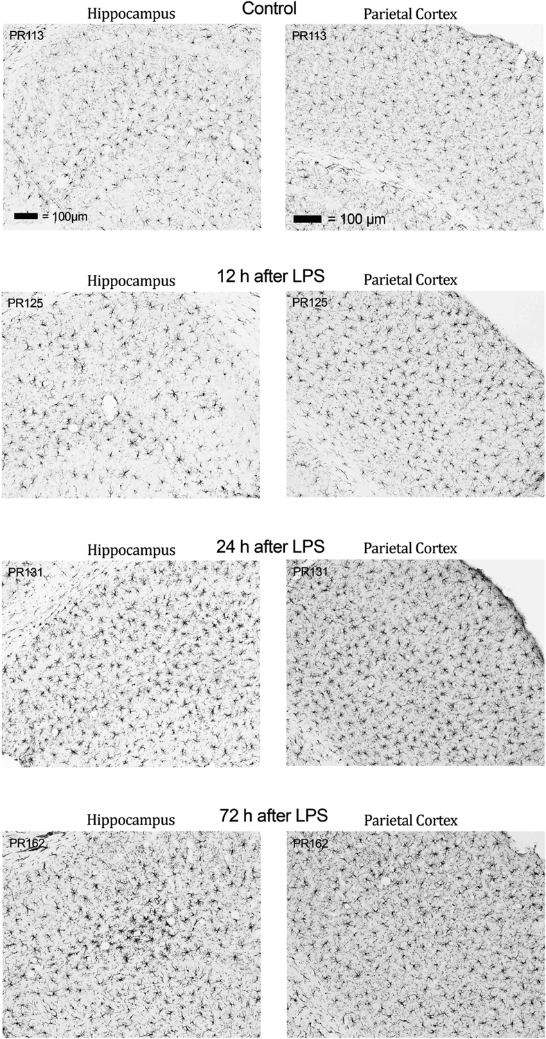
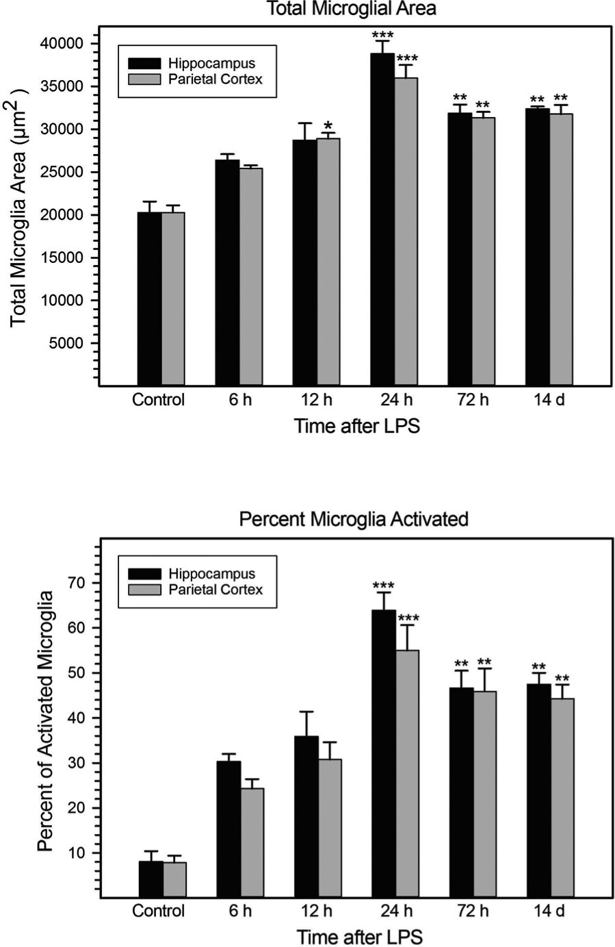
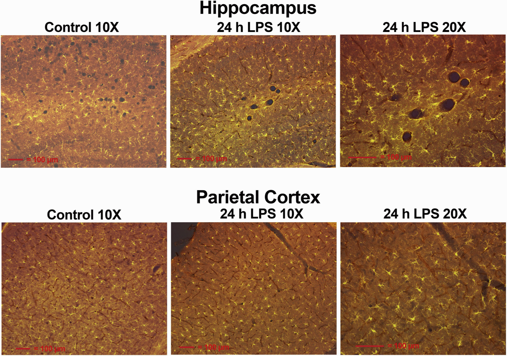
Description:Bacterial cell wall endotoxins, i.e. lipopolysaccharides (LPS), are some of the original compounds shown to evoke the classic signs of systemic inflammation/innate immune response and neuroinflammation. The term neuroinflammation often is used to infer the elaboration of proinflammatory mediators by microglia elicited by neuronal targeted activity. However, it also is possible that the microglia are responding to vasculature through several signaling mechanisms. Microglial activation relative to the vasculature in the hippocampus and parietal cortex was determined after an acute exposure of a single subcutaneous injection of 2 mg/kg LPS. Antibodies to allograft inflammatory factor (Aif1, a.k.a. Iba1) were used to track and quantify morphological changes in microglia. Immunostaining of platelet/endothelial cell adhesion molecule 1 (Pecam1, a.k.a. Cd31) was used to visualize vasculature in the forebrain and glial acidic fibrillary protein (GFAP) to visualize astrocytes. Neuroinflammation and other aspects of neurotoxicity were evaluated histologically at 3 h, 6 h, 12 h, 24 h, 3 d and 14 d following LPS exposure. LPS did not cause neurodegeneration as determined by Fluoro Jade C labeling. Also, there were no signs of mouse IgG leakage from brain vasculature due to LPS. Some changes in microglia size occurred at 6 h, but by 12 h microglial activation had begun with the combined soma and proximal processes size increasing significantly (1.5-fold). At 24 h, almost all the microglia soma and proximal processes in the hippocampus, parietal cortex, and thalamus were closely associated with the vasculature and had increased almost 2.0-fold in size. In many areas where microglia were juxtaposed to vasculature, astrocytic endfeet appeared to be displaced. The microglial activation had subsided slightly by 3 d with microglial size 1.6-fold that of control. We hypothesize that acute LPS activation can result in vascular mediated microglial responses through several mechanisms: 1) binding to Cd14 and Tlr4 receptors on microglia processes residing on vasculature; 2) damaging vasculature and causing the release of cytokines; and 3) possibly astrocytic endfeet damage resulting in cytokine release. These acute responses may serve as an adaptive mechanism to exposure to circulating LPS where the microglia surround the vasculature. This could further prevent the pathogen(s) circulating in blood from entering the brain. However, diverting microglial interactions away from synaptic remodeling and other types of microglial interactions with neurons may have adverse effects on neuronal function.
-
Subjects:
-
Source:
-
Pubmed ID:32014511
-
Pubmed Central ID:PMC7294247
-
Document Type:
-
Collection(s):
-
Main Document Checksum:
-
Download URL:
-
File Type:
-

gif 
jpeg 
gif 
jpeg 
gif 
jpeg 
xml 
pdf 
doc 
docx 
gif 
xlsx 
jpeg 
gif 
jpeg 
gif 
jpeg 
gif 
jpeg
 [PDF-2.39 MB]
Details:
Supporting Files


More +
You May Also Like

 [PDF - 687.57 KB]
 Email
CDC-INFO
Email
CDC-INFO