i
Pulmonary fibrotic response to aspiration of multi-walled carbon nanotubes
-
Jul 22 2011
Source: Part Fibre Toxicol. 2011; 8:21.
 [PDF-3.75 MB]
Details:
-
Alternative Title:Part Fibre Toxicol
-
Personal Author:
-
Description:Background
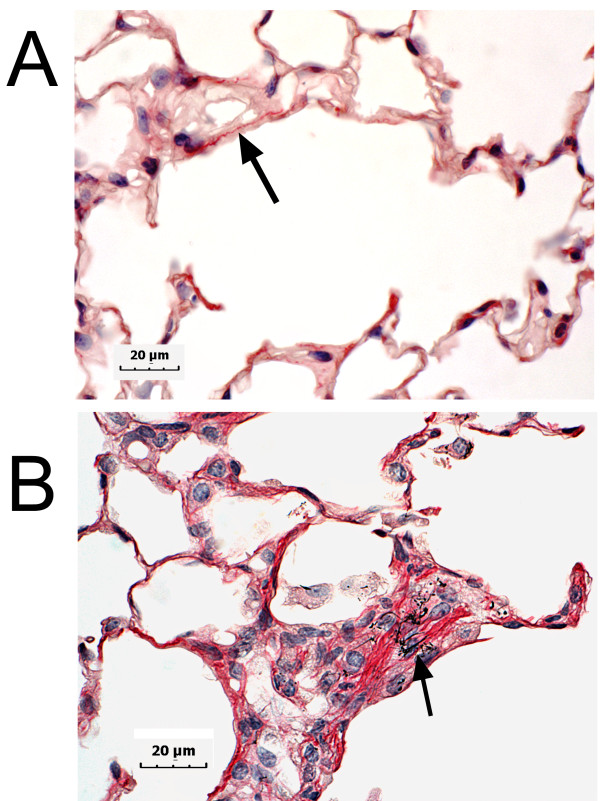
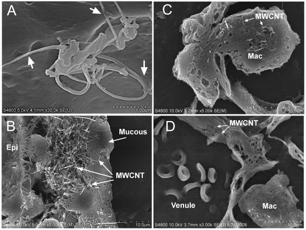
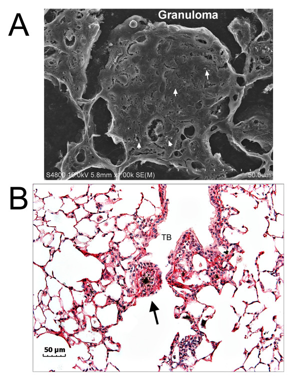
Multi-walled carbon nanotubes (MWCNTs) are new manufactured nanomaterials with a wide spectrum of commercial applications. To address the hypothesis that MWCNTs cause persistent pulmonary pathology, C57BL/6J mice were exposed by pharyngeal aspiration to 10, 20, 40 or 80 μg of MWCNTs (mean dimensions of 3.9 μm × 49 nm) or vehicle. Lungs were preserved at 1, 7, 28 and 56 days post- exposure to determine the potential regions and target cells for impact by MWCNT lung burden. Morphometric measurement of Sirius Red staining was used to assess the connective tissue response.
Results
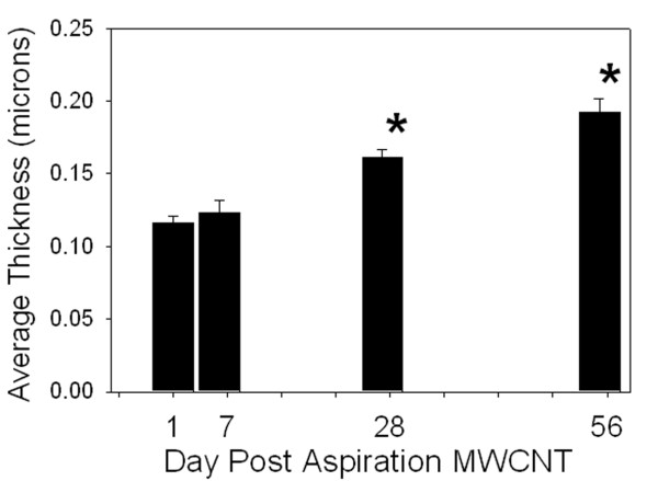
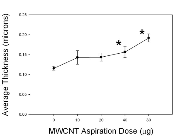
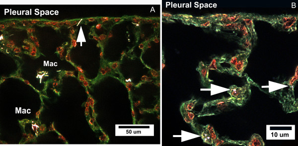
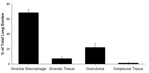
At 56 days post-exposure, 68.7 ± 3.9, 7.5 ± 1.9 and 22.0 ± 5.1 percent (mean ± SE, N = 8) of the MWCNT lung burden were in alveolar macrophages, alveolar tissue and granulomatous lesions, respectively. The subpleural tissues contained 1.6% of the MWCNT lung burden. No MWCNTs were found in the airways at 7, 28 or 56 days after aspiration The connective tissue in the alveolar interstitium demonstrated a progressive increase in thickness over time in the 80 μg exposure group (0.12 ± 0.01, 0.12 ± 0.01, 0.16 ± 0.01 and 0.19 ± 0.01 μm for 1, 7, 28 and 56 days post-exposure (mean ± SE, N = 8)). Dose-response determined at 56 days post-exposure for the average thickness of connective tissue in alveolar septa was 0.11 ± 0.01, 0.14 ± .02, 0.14 ± 0.01, 0.16 ± 0.01 and 0.19 ± 0.01 μm (mean ± SE, N = 8) for vehicle, 10, 20, 40 and 80 μg dose groups, respectively.
Conclusions
The distribution of lung burden was predominately within alveolar macrophages with approximately 8% delivery to the alveolar septa, and a smaller but potentially significant burden to the subpleural tissues. Despite the relatively low fraction of the lung burden being delivered to the alveolar tissue, the average thickness of connective tissue in the alveolar septa was increased over vehicle control by 45% in the 40 μg and 73% in the 80 μg exposure groups. The results demonstrate that MWCNTs have the potential to produce a progressive, fibrotic response in the alveolar tissues of the lungs. However, the increases in connective tissue per μg dose of MWCNTs to the interstitium are significantly less than those previously found for single-walled carbon nanotubes (SWCNTs).
-
Subjects:
-
Source:
-
Document Type:
-
Collection(s):
-
Main Document Checksum:
-
Download URL:
-
File Type:
Supporting Files
-

gif 
jpeg 
gif 
jpeg 
gif 
jpeg 
txt
txt 
gif 
jpeg 
gif 
jpeg 
gif 
jpeg 
gif 
jpeg


More +


 Email
CDC-INFO
Email
CDC-INFO